Ankle Anatomy Basics
Understanding ankle anatomy and the structures that are most commonly injured will help you prevent and treat common ankle sprains and injuries. Did you know that ankle injuries are the single most common injury suffered during sports participation?
We often overlook ankle injuries, chalking them up to just being part of the game. A good understanding of the anatomy and structures that can be injured can help you learn how to treat your ankle injury and prevent future ankle injuries.
Ankle and Foot Bones
The ankle joint is made up of 3 bones that articulate (touch) with each
other. The tibia, fibula, and talus. The tibia is the weight
bearing bone of the lower leg...also known as
the "shin bone". The fibula is the smaller low leg bone, on the outside
of your leg. Both the tibia and fibula extend down to the ankle, and
their ends create the "ankle bones" on the inside and outside of the
ankle, called the malleoli.
The talus sits between the tibia and the fibula, and creates the ankle
"mortise". This joint acts like a hinge joint, allowing you to move
your foot up and down.
Other bones important in ankle anatomy include the calcaneus (heel
bone), the tarsals, metatarsals, and phalanges (toes).
Take the Ankle Anatomy Video Tour

Moving further down the foot are the tarsals - five
bones that form the mid-section of your foot. Each of these bones is
connected to each other with small ligaments. The tarsals
make up the midfoot, and part of the arch of the foot. There
is not a
lot of movement between these bones, however, they do need to be able
to give and take as you bear weight on the leg.
Following the foot towards the toes, next
comes the metatarsals, five bones that make up the end of your foot,
just before the toes. This is referred to as the forefoot. The
metatarsals are the most commonly injured bones in the ankle. Most fractures and stress fractures occur here.
Last, but not least, the
final bones are the
phalanges - better known as the toes. Each toe is
made up of three
bones, except for the big toe, which only has two. Toes
are commonly
injured in sports, however, these injuries are usually minor.
Foot and Ankle Anatomy - Ligaments
Ligaments are commonly sprained at the ankle, with ankle sprains being the most common sports injury. There are several ligaments of importance in the ankle.
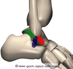
The lateral ankle
is comprised of three different ligaments. In the picture on
the
right, the
Anterior Talo-fibular Ligament
is shown in red. This is the most
commonly injured ligament. It runs between the talus and the fibula.
The Calcaneal
Fibular Ligament
is shown in blue. It runs between the
fibula and the calcaneus.
The Posterior
Talo-fibular Ligament is shown in green and runs between
the fibula and the posterior talus.
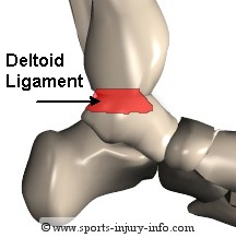
The medial side of the ankle is
comprised of one large ligament that spans the entire end of the tibia.
This ligament runs between the tibia and the navicular and calcaneus. It
has several areas of thickening, or bands, but is considered
one ligament.
It is not commonly injured with
ankle sprains, as the vast majority of sprains occur by rolling over
the outside of the ankle.
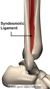
You may have heard sports announcers talk about a "high ankle sprain". This type of injury involves the
sydesmotic ligament, which runs between the tibia and the fibula above
the ankle joint. Injury of this ligament is not common. This ligament is sprained when the
fibula and the tibia are seperated or pushed apart. It can occur with
forced dorsiflexion of the ankle, or with with a very forceful landing.
The
majority of ankle sprains do not involve this ligament, however, those
that do can take an extra long time to heal because of the stress
placed
on this ligament during weight bearing activities.
Foot ligaments include all of the ligaments that connect the tarsal bones, as well as the ligaments and joint capsules of the metatarsals and phalanges. Ligaments are responsible for maintaining the arch of the foot, especially at the tarsal bones in the midfoot. The plantar fascia is not a ligament, but does help to maintain the arch, and may become inflammed with plantar fasciitis.
Ankle and Foot Anatomy - Muscles
Muscles that originate (or start) at the low leg insert (or attach) to the bones in the ankle and foot and help produce motion at the ankle and foot. Muscles also help to provide stability during activities.
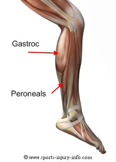
The calf muscles, the gastroc and soleus,
allow for pointing of the foot, as well as lifting up on your
"tip-toes".
The peroneal muscles (also called fibularis muscles), on the outside of your leg,
help to keep you stable during activity, and also help turn the foot
out (called eversion). Other smaller muscles on the front and inside of
the leg help to flex and extend your foot and toes. These include the anterior tibialis, posterior tibialis, and flexor digitorum, extensor and flexor hallicus, and extensor digitorum longus and brevis.
The majority of the foot muscles are small, intrinsic muscles that run along the underside of the foot. They help to move the toes, and to maintain the arch of the foot. Most of the muscles in the foot that are injured with sports originate from the lower leg and cross the ankle.
Ankle Anatomy - Tendons
Tendons are also a common site for sports injury. All of the
muscles of the lower leg attach to the ankle and foot through tendons. A common injury that occurs due to tissue overstress is tendonitis. The
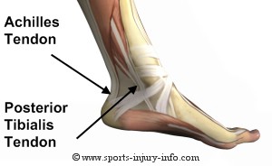
medial side of the ankle is home to several different tendons. The most
commonly injured of these is the posterior tibialis tendon. It runs
behind the medial malleolus (ankle bone) and attaches at the mid-foot.
This
tendon helps to maintain the arch of the foot, and to control pronation.
Also
pictured is the achilles tendon, which attaches to the calcaneus. This
is the insertion for the calf muscles, and is a very common site for achilles tendonitis.
Also
on the medial side of the ankle are the flexor hallicus longus and
flexor digitorum. These tendons run along the medial malleolus with the
posterior tibialis, and attach to the great toe and the lesser toes
respectively.
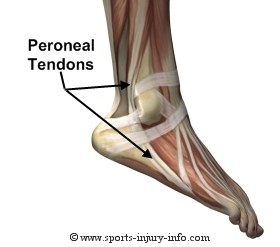
The lateral ankle tendons include the peroneals, or fibularis tendons. There are three peroneal tendons, and they all run along the outside of
the ankle behind the lateral malleolus. They attach at the base of the 5th metatarsal, and also on the
bottom of the foot.
Peroneal tendonitis is a common
problem with sports activities. These tendons can also be strained with
ankle sprains. With the forceful
turning of the ankle, not only are the ligaments injured, but also the
tendons.
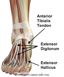
The tendon that runs down the front of the leg and attaches to
the midfoot tarsal bones is the anterior tibialis. It helps to pull the foot towards the body,
and to control motion during activity. This is often the muscle and tendon that is irritated with shin splints.
The other
tendons on the
anterior side of the ankle, or the top of the foot include the extensor
digitorum and the extensor hallicus tendons. The extensor digitorum
attaches to the lesser toes, while the extensor hallicus attaches to
the great toe.
If you are currently suffering from achilles tendonitis, plantar
faciitis, ankle, or foot pain, you could benefit from a comprehensive
foot and ankle
strengthening program. Foot Pain Solutions
is exactly what you need to help eliminate
your foot, ankle, and low leg pain. Great for prevention too, Foot Pain
Solutions can guide you to finally getting rid of your chronic foot and
ankle pain.
All of the muscles, bones, and ligaments work together to keep your ankle and foot in tip top shape. Understanding ankle anatomy is the first key to prevention and treatment of ankle injuries. You can learn more about ankle anatomy with our Ankle Video Tour. I'll walk you through all of the ankle anatomy step by step.
SII › Ankle Anatomy
Recent Articles
-
Common sports injuries - Straight forward information from Sports Injury Info
Nov 25, 15 05:44 PM
 Learn more about common sports injuries. From ACL tears to ankle sprains, we include the information you need to identify your injury and determine the best course of action.
 -
Stories of Multiple ACL Tears
Sep 25, 13 05:03 PM
 Read stories about visitors who suffered multiple ACL tears, how they recovered, and tips for prevention.
 -
Australia - Christian's soccer acl recovery age 13
Apr 24, 12 12:47 AM
Hi guys, I'm now nearly 6 months into my recovery. I had a FFA soccer scholarship and things were looking good until I injured myself on the 2nd day of
Subscribe to
"The Athletic Trainer's Corner"